What I study
Jul 19, 2024
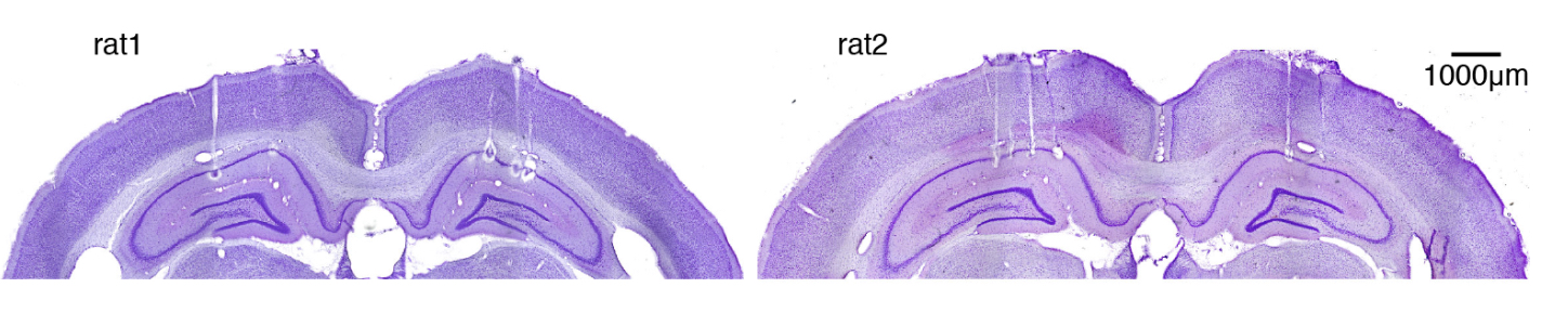
The brain carries out many different tasks; there are countless number of cells (neurons and neuron maintainence cells), cell types in various brain regions. However, if you take a cross section of the brain and stain them to locate cells; you’d see a couple dense strips. These dense strips of cells lie in the brain region hippocampus. Not only does the hippocampus have a striking dense collection of cells (prominent in the way it looks), the region is central to memory, navigation, and relational thinking. I study the main output part of the hippocampus (prominent in its computation power).
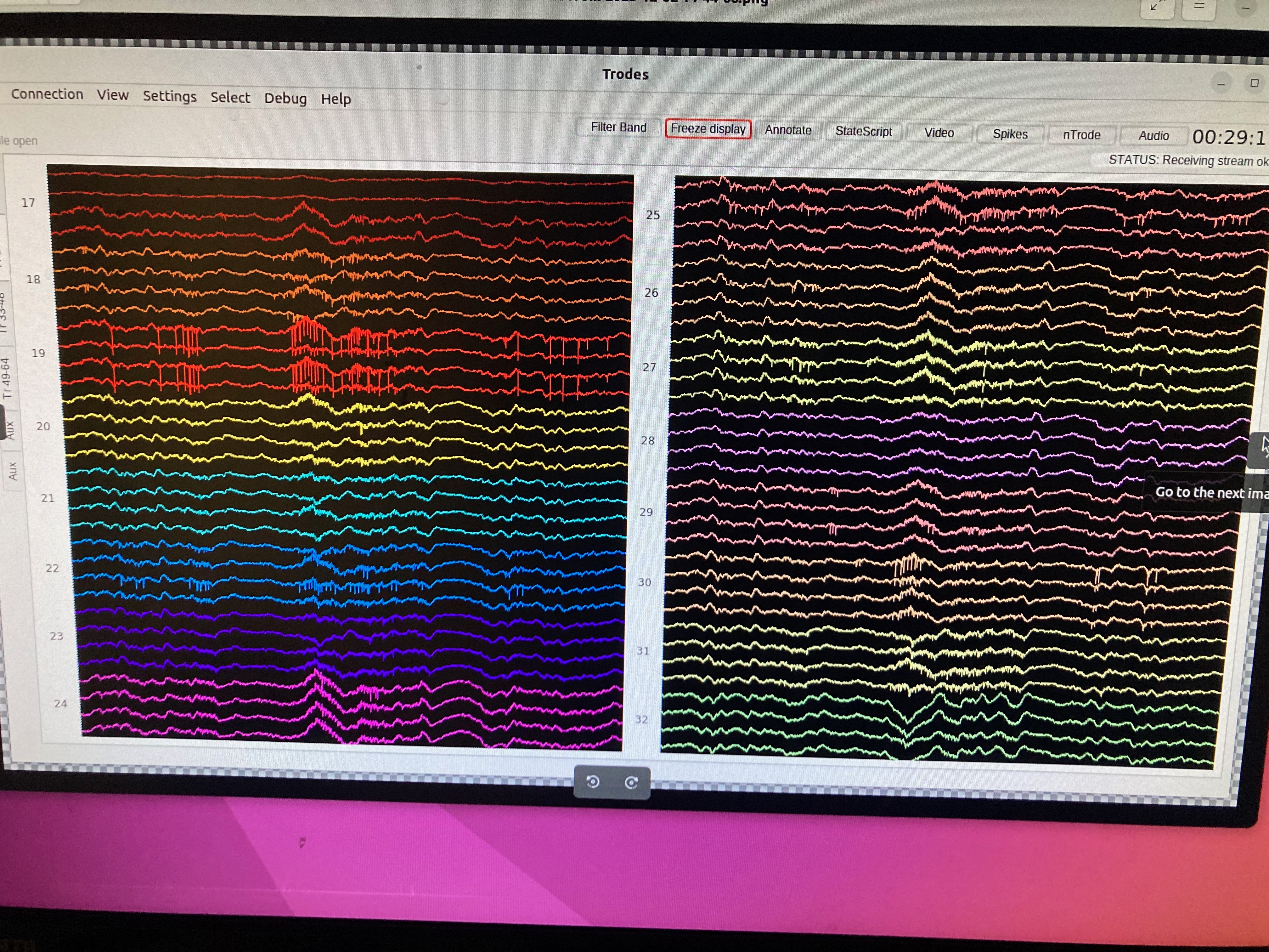
The cover picture shows what the activities in the CA1 region of the hippocampus look like when a lot of cells fire around the same time — talking about 10s of ms. These strong deflections are thought to be the key driver for memory consolidation.
How far are we to understand this? We know a lot and very little at the same time. Stay tuned.